| 產(chǎn)品編號 | bsm-33036M |
| 英文名稱 | Mouse Anti-beta-Actin (Loading Control) antibody |
| 中文名稱 | β-肌動蛋白/β-Actin(內(nèi)參)單克隆抗體 |
| 別 名 | Beta Actin; beta-Actin; ACTB; Actin cytoplasmic 1; Actin, beta; Beta actin; beta cytoskeletal actin;A X actin like protein; ACTB; Actin cytoplasmic 1; alpha sarcomeric Actin; Actx; Beta cytoskeletal actin; Melanoma X actin; PS1TP5BP1; ACTB_HUMAN. |
 | Specific References (85) | bsm-33036M has been referenced in 85 publications. |
| 產(chǎn)品類型 | 內(nèi)參抗體 |
| 研究領(lǐng)域 | 腫瘤 細胞生物 信號轉(zhuǎn)導(dǎo) 細胞骨架 |
| 抗體來源 | Mouse |
| 克隆類型 | Monoclonal |
| 克 隆 號 | 1A2 |
| 交叉反應(yīng) | Human,Mouse,Rat (predicted: Chicken,Dog,Pig,Cow,Sheep,Fish,GuineaPig,Hamster,Cat) |
| 產(chǎn)品應(yīng)用 | WB=1:5000-20000, IHC-P=1:200-1000, IHC-F=1:200-1000, ICC=1:100, IF=1:200-500, Flow-Cyt=1ug/Test, ELISA=1:5000-10000 not yet tested in other applications. optimal dilutions/concentrations should be determined by the end user. |
| 理論分子量 | 42kDa |
| 細胞定位 | 細胞漿 |
| 性 狀 | Liquid |
| 濃 度 | 1mg/ml |
| 免 疫 原 | Synthetic MAP peptide derived from human beta-Actin |
| 亞 型 | IgG |
| 純化方法 | affinity purified by Protein G |
| 緩 沖 液 | 0.01M TBS(pH7.4) with 1% BSA, 0.03% Proclin300 and 50% Glycerol. |
| 保存條件 | Shipped at 4℃. Store at -20 °C for one year. Avoid repeated freeze/thaw cycles. |
| 注意事項 | This product as supplied is intended for research use only, not for use in human, therapeutic or diagnostic applications. |
| PubMed | PubMed |
| 產(chǎn)品介紹 | Loading Control This gene encodes one of six different actin proteins. Actins are highly conserved proteins that are involved in cell motility, structure, and integrity. This actin is a major constituent of the contractile apparatus and one of the two nonmuscle cytoskeletal actins. [provided by RefSeq, Jul 2008]. Function: Actins are highly conserved proteins that are involved in various types of cell motility and are ubiquitously expressed in all eukaryotic cells. Subunit: Polymerization of globular actin (G-actin) leads to a structural filament (F-actin) in the form of a two-stranded helix. Each actin can bind to 4 others. Identified in a mRNP granule complex, at least composed of ACTB, ACTN4, DHX9, ERG, HNRNPA1, HNRNPA2B1, HNRNPAB, HNRNPD, HNRNPL, HNRNPR, HNRNPU, HSPA1, HSPA8, IGF2BP1, ILF2, ILF3, NCBP1, NCL, PABPC1, PABPC4, PABPN1, RPLP0, RPS3, RPS3A, RPS4X, RPS8, RPS9, SYNCRIP, TROVE2, YBX1 and untranslated mRNAs. Component of the BAF complex, which includes at least actin (ACTB), ARID1A, ARID1B/BAF250, SMARCA2, SMARCA4/BRG1, ACTL6A/BAF53, ACTL6B/BAF53B, SMARCE1/BAF57 SMARCC1/BAF155, SMARCC2/BAF170, SMARCB1/SNF5/INI1, and one or more of SMARCD1/BAF60A, SMARCD2/BAF60B, or SMARCD3/BAF60C. In muscle cells, the BAF complex also contains DPF3. Found in a complex with XPO6, Ran, ACTB and PFN1. Component of the MLL5-L complex, at least composed of MLL5, STK38, PPP1CA, PPP1CB, PPP1CC, HCFC1, ACTB and OGT. Interacts with XPO6 and EMD. Interacts with ERBB2. Subcellular Location: Cytoplasm. cytoskeleton. Tissue Specificity: Ubiquitously expressed in all eukaryotic cells. Post-translational modifications: ISGylated. Oxidation of Met-44 by MICALs (MICAL1, MICAL2 or MICAL3) to form methionine sulfoxide promotes actin filament depolymerization. Methionine sulfoxide is produced stereospecifically, but it is not known whether the (S)-S-oxide or the (R)-S-oxide is produced. DISEASE: Defects in ACTA1 are the cause of nemaline myopathy type 3 (NEM3) [MIM:161800]. A form of nemaline myopathy. Nemaline myopathies are muscular disorders characterized by muscle weakness of varying severity and onset, and abnormal thread-or rod-like structures in muscle fibers on histologic examination. The phenotype at histological level is variable. Some patients present areas devoid of oxidative activity containg (cores) within myofibers. Core lesions are unstructured and poorly circumscribed. Defects in ACTA1 are a cause of myopathy congenital with excess of thin myofilaments (MPCETM) [MIM:161800]. A congenital muscular disorder characterized at histological level by areas of sarcoplasm devoid of normal myofibrils and mitochondria, and replaced with dense masses of thin filaments. Central cores, rods, ragged red fibers, and necrosis are absent. Similarity: Belongs to the actin family. SWISS: P60709 Gene ID: 60 Database links: Entrez Gene: 396526 Chicken Entrez Gene: 60 Human Entrez Gene: 11461 Mouse Entrez Gene: 100009272 Rabbit Omim: 102630 Human SwissProt: P60706 Chicken SwissProt: P60708 Horse SwissProt: P60709 Human SwissProt: P60710 Mouse SwissProt: P29751 Rabbit SwissProt: P60713 Sheep Unigene: 520640 Human Unigene: 708120 Human Unigene: 727576 Human Unigene: 328431 Mouse Unigene: 391967 Mouse Unigene: 94978 Rat |
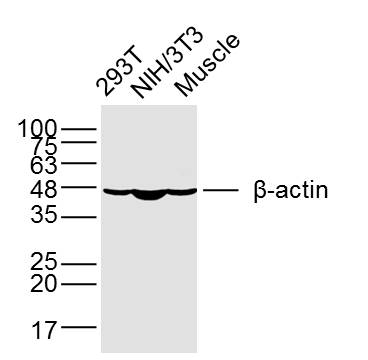
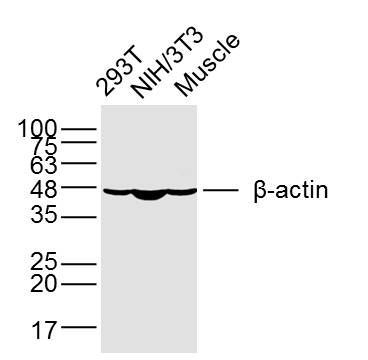
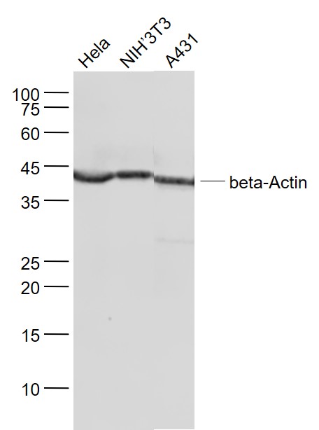
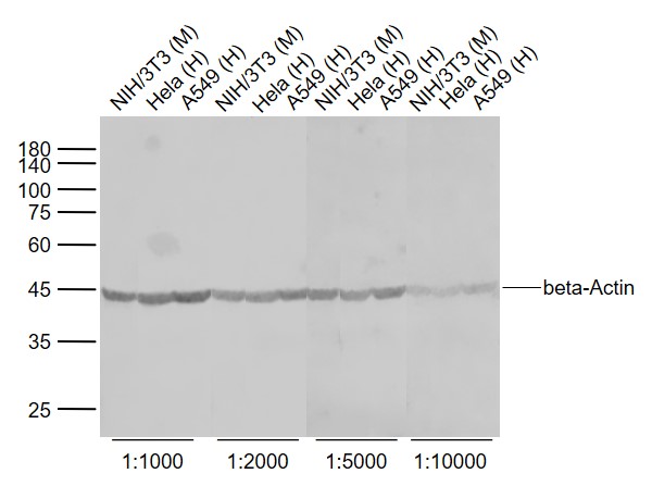
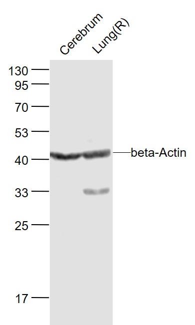
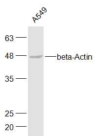
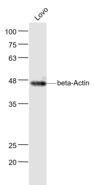
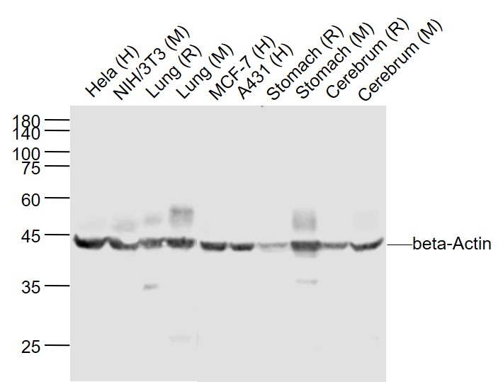
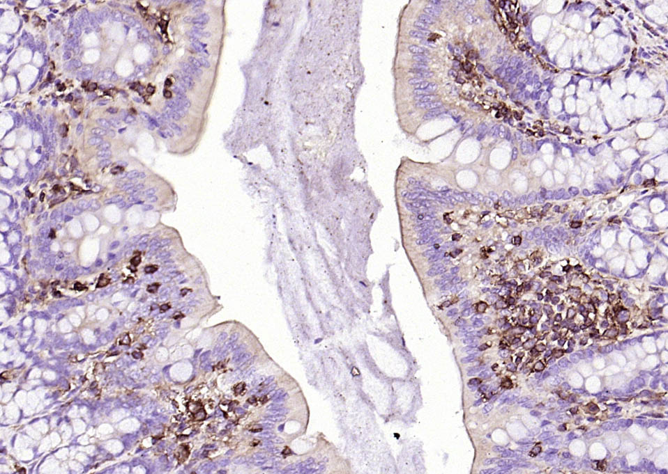
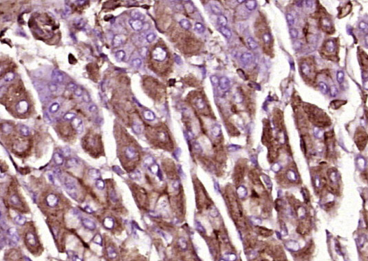
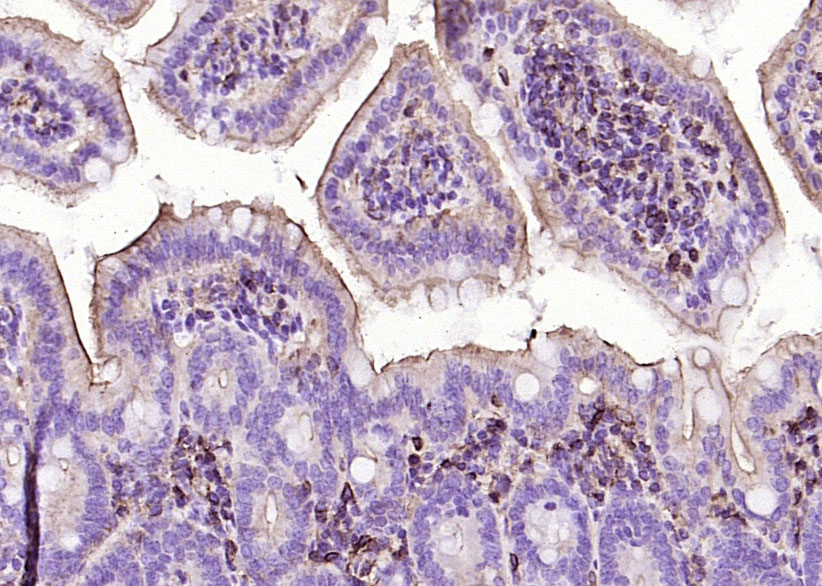
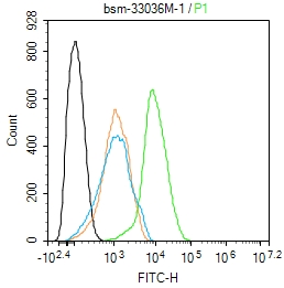
| 產(chǎn)品圖片 | Sample: 293T Cell (Human) Lysate at 40 ug NIH/3T3 Cell (Mouse) Lysate at 40 ug Muscle (Mouse) Lysate at 40 ug Primary: Anti-β-Actin (bsm-33036M) at 1/5000 dilution Secondary: IRDye800CW Goat Anti-Mouse IgG at 1/20000 dilution Predicted band size: 42 kD Observed band size: 42 kD Sample: Hela(Human) Cell Lysate at 30 ug NIH/3T3(Mouse) Cell Lysate at 30 ug A431(Human) Cell Lysate at 30 ug Primary: Anti-beta-Actin (bsm-33036M) at 1/1000 dilution Secondary: IRDye800CW Goat Anti-Mouse IgG at 1/20000 dilution Predicted band size: 42 kD Observed band size: 42 kD Sample: NIH/3T3 (Mouse) Cell Lysate at 30 ug Hela (Human) Cell Lysate at 30 ug A549 (Human) Cell Lysate at 30 ug Primary: Anti-beta-Actin (bsm-33036M) at 1/1000~1/10000 dilution Secondary: IRDye800CW Goat Anti-Mouse IgG at 1/20000 dilution Predicted band size: 42 kD Observed band size: 42 kD Sample: Cerebrum(Mouse) Lysate at 40 ug Lung(Rat) Lysate at 40 ug Primary: Anti-beta-Actin (bsm-33036M) at 1/1000 dilution Secondary: IRDye800CW Goat Anti-Rabbit IgG at 1/20000 dilution Predicted band size: 42 kD Observed band size: 42 kD Sample: A549(Human) Cell Lysate at 30 ug Primary: Anti-beta-Actin (bsm-33036M) at 1/500 dilution Secondary: IRDye800CW Goat Anti-Mouse IgG at 1/20000 dilution Predicted band size: 42 kD Observed band size: 42 kD Sample: Lovo(Human) Cell Lysate at 30 ug Primary: Anti- beta-Actin (bsm-33036M) at 1/500 dilution Secondary: IRDye800CW Goat Anti-Mouse IgG at 1/20000 dilution Predicted band size: 42 kD Observed band size: 46 kD Sample: Lane 1: Hela (Human) Cell Lysate at 30 ug Lane 2: NIH/3T3 (Mouse) Cell Lysate at 30 ug Lane 3: Lung (Rat) Lysate at 40 ug Lane 4: Lung (Mouse) Lysate at 40 ug Lane 5: MCF-7 (Human) Cell Lysate at 30 ug Lane 6: A431 (Human) Cell Lysate at 30 ug Lane 7: Stomach (Rat) Lysate at 40 ug Lane 8: Stomach (Mouse) Lysate at 40 ug Lane 9: Cerebrum (Rat) Lysate at 40 ug Lane 10: Cerebrum (Mouse) Lysate at 40 ug Primary: Anti-beta-Actin (bsm-33036M) at 1/1000 dilution Secondary: IRDye800CW Goat Anti-Mouse IgG at 1/20000 dilution Predicted band size: 42 kD Observed band size: 42 kD Paraformaldehyde-fixed, paraffin embedded (rat colon); Antigen retrieval by boiling in sodium citrate buffer (pH6.0) for 15min; Block endogenous peroxidase by 3% hydrogen peroxide for 20 minutes; Blocking buffer (normal goat serum) at 37°C for 30min; Antibody incubation with (beta-Actin (Loading Control)) Monoclonal Antibody, Unconjugated (bsm-33036M) at 1:200 overnight at 4°C, followed by operating according to SP Kit(Mouse)(sp-0024) instructionsand DAB staining. Paraformaldehyde-fixed, paraffin embedded (mouse stomach tissue); Antigen retrieval by boiling in sodium citrate buffer (pH6.0) for 15min; Block endogenous peroxidase by 3% hydrogen peroxide for 20 minutes; Blocking buffer (normal goat serum) at 37°C for 30min; Antibody incubation with (beta-Actin) Monoclonal Antibody, Unconjugated (ascites of bsm-33036M) at 1:2000 overnight at 4°C, followed by a conjugated secondary (sp-0024) for 20 minutes and DAB staining. Paraformaldehyde-fixed, paraffin embedded (mouse colon); Antigen retrieval by boiling in sodium citrate buffer (pH6.0) for 15min; Block endogenous peroxidase by 3% hydrogen peroxide for 20 minutes; Blocking buffer (normal goat serum) at 37°C for 30min; Antibody incubation with (beta-Actin (Loading Control)) Monoclonal Antibody, Unconjugated (bsm-33036M) at 1:200 overnight at 4°C, followed by operating according to SP Kit(Mouse)(sp-0024) instructionsand DAB staining. Blank control:Hela. Primary Antibody (green line): Mouse Anti-beta-Actin antibody (bsm-33036M) Dilution: 1ug/Test; Secondary Antibody : Goat anti-Mouse IgG-FITC Dilution: 0.5ug/Test. Protocol The cells were fixed with 4% PFA (10min at room temperature)and then permeabilized with 90% ice-cold methanol for 20 min at -20℃.The cells were then incubated in 5%BSA to block non-specific protein-protein interactions for 30 min at room temperature .Cells stained with Primary Antibody for 30 min at room temperature. The secondary antibody used for 40 min at room temperature. Acquisition of 20,000 events was performed. |


