| 產(chǎn)品編號 | bsm-52056R |
| 英文名稱 | Rabbit Anti-Cytokeratin 16 antibody |
| 中文名稱 | 細(xì)胞角蛋白16重組兔單抗 |
| 別 名 | Cytokeratin 16; CK 16; Focal non epidermolytic palmoplantar keratoderma; K 16; K16; K1CP; CK-16; CK16; Cytokeratin-16; FNEPPK; K1C16_HUMAN; Keratin; Keratin-16; type I cytoskeletal 16; Keratin 16; Keratin type I cytoskeletal 16; Keratin16; KRT 16; KRT16; KRT16A; NEPPK. |
| 研究領(lǐng)域 | 腫瘤 信號轉(zhuǎn)導(dǎo) 細(xì)胞骨架 |
| 抗體來源 | Rabbit |
| 克隆類型 | Recombinant |
| 克 隆 號 | 1A4 |
| 交叉反應(yīng) | Human,Mouse,Rat |
| 產(chǎn)品應(yīng)用 | WB=1:500-2000, IHC-P=1:100-500, IHC-F=1:400-800, ICC=1:50-200, IF=1:50-200 not yet tested in other applications. optimal dilutions/concentrations should be determined by the end user. |
| 理論分子量 | 51kDa |
| 細(xì)胞定位 | 細(xì)胞漿 |
| 性 狀 | Liquid |
| 濃 度 | 1mg/ml |
| 免 疫 原 | Recombinant human Cytokeratin 16 protein (1-100aa) |
| 亞 型 | IgG |
| 純化方法 | affinity purified by Protein A |
| 緩 沖 液 | 0.01M TBS(pH7.4) with 1% BSA, 0.03% Proclin300 and 50% Glycerol. |
| 保存條件 | Shipped at 4℃. Store at -20 °C for one year. Avoid repeated freeze/thaw cycles. |
| 注意事項(xiàng) | This product as supplied is intended for research use only, not for use in human, therapeutic or diagnostic applications. |
| PubMed | PubMed |
| 產(chǎn)品介紹 | The protein encoded by this gene is a member of the keratin gene family. The keratins are intermediate filament proteins responsible for the structural integrity of epithelial cells and are subdivided into cytokeratins and hair keratins. Most of the type I cytokeratins consist of acidic proteins which are arranged in pairs of heterotypic keratin chains and are clustered in a region of chromosome 17q12-q21. This keratin has been coexpressed with keratin 14 in a number of epithelial tissues, including esophagus, tongue, and hair follicles. Mutations in this gene are associated with type 1 pachyonychia congenita, non-epidermolytic palmoplantar keratoderma and unilateral palmoplantar verrucous nevus. [provided by RefSeq, Jul 2008]. Subunit: Heterodimer of a type I and a type II keratin. KRT16 associates with KRT6 isomers. Interacts with TCHP. Interacts with TRADD. Tissue Specificity: Expressed in the hair follicle, nail bed and in mucosal stratified squamous epithelia and, suprabasally, in oral epithelium and palmoplantar epidermis. Also found in luminal cells of sweat and mammary gland ducts. DISEASE: Pachyonychia congenita 1 (PC1) [MIM:167200]: An autosomal dominant ectodermal dysplasia characterized by hypertrophic nail dystrophy resulting in onchyogryposis (thickening and increase in curvature of the nail), palmoplantar keratoderma, follicular hyperkeratosis, and oral leukokeratosis. Hyperhidrosis of the hands and feet is usually present. Note=The disease is caused by mutations affecting the gene represented in this entry. Keratoderma, palmoplantar, non-epidermolytic, focal (FNEPPK) [MIM:613000]: A dermatological disorder characterized by non-epidermolytic palmoplantar keratoderma limited to the pressure points on the balls of the feet, with later mild involvement on the palms. Oral, genital and follicular keratotic lesions are often present. Note=The disease is caused by mutations affecting the gene represented in this entry. Unilateral palmoplantar verrucous nevus (UPVN) [MIM:144200]: UPVN is characterized by a localized epidermolytic hyperkeratosis in parts of the right palm and the right sole, following the lines of Blaschko. Note=The disease is caused by mutations affecting the gene represented in this entry. Note=KRT16 and KRT17 are coexpressed only in pathological situations such as metaplasias and carcinomas of the uterine cervix and in psoriasis vulgaris. Similarity: Belongs to the intermediate filament family. SWISS: P08779 Gene ID: 3868 Database links: Entrez Gene: 3868 Human Omim: 148067 Human SwissProt: P08779 Human Unigene: 655160 Human |
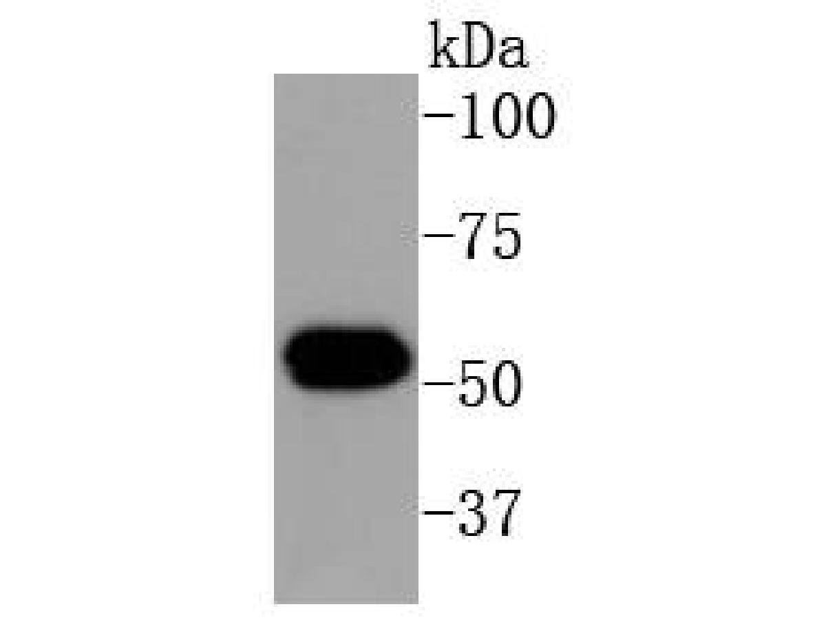
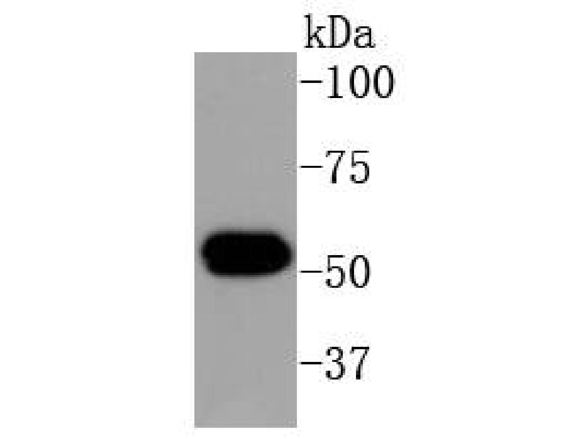
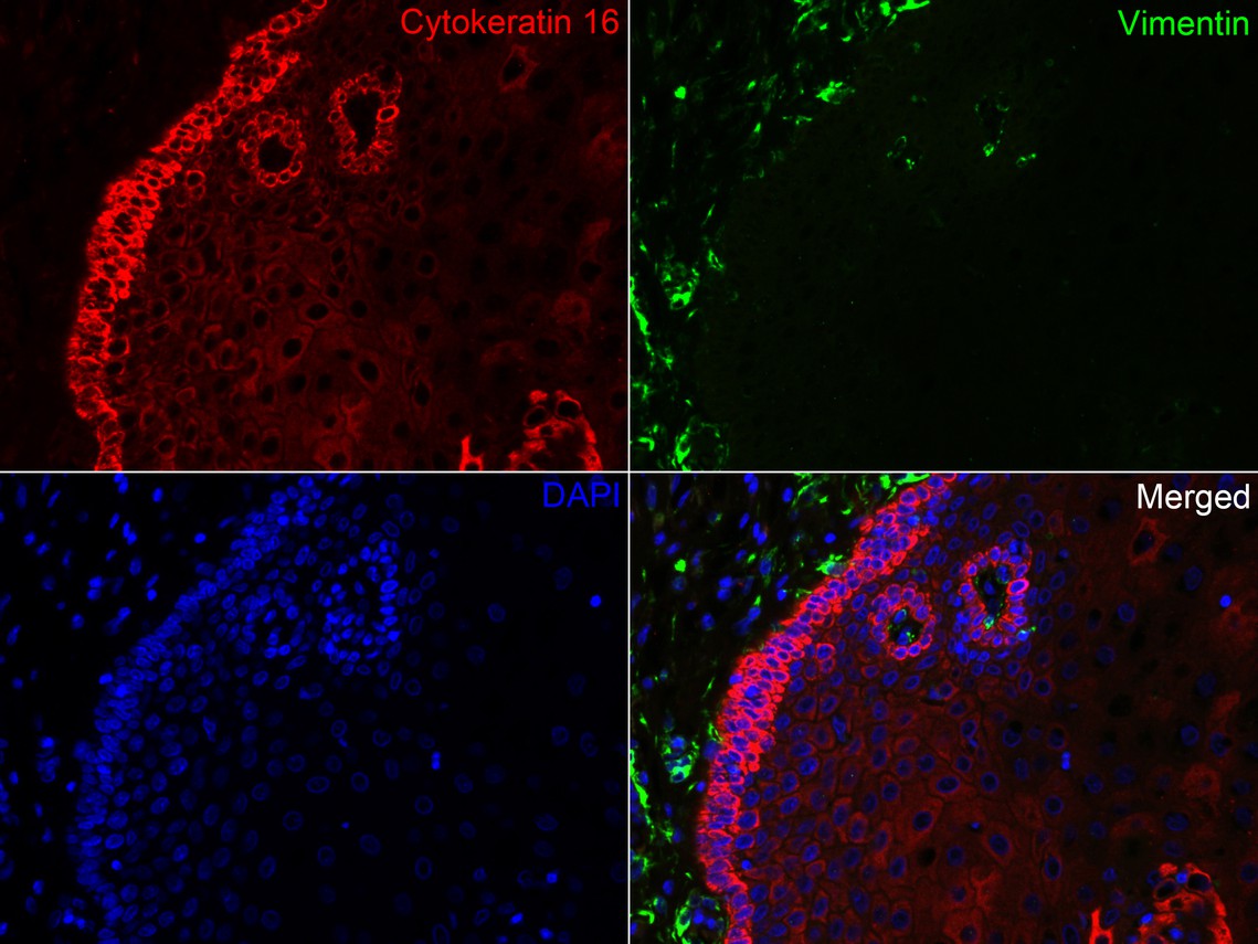
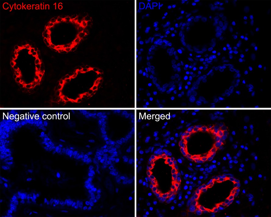
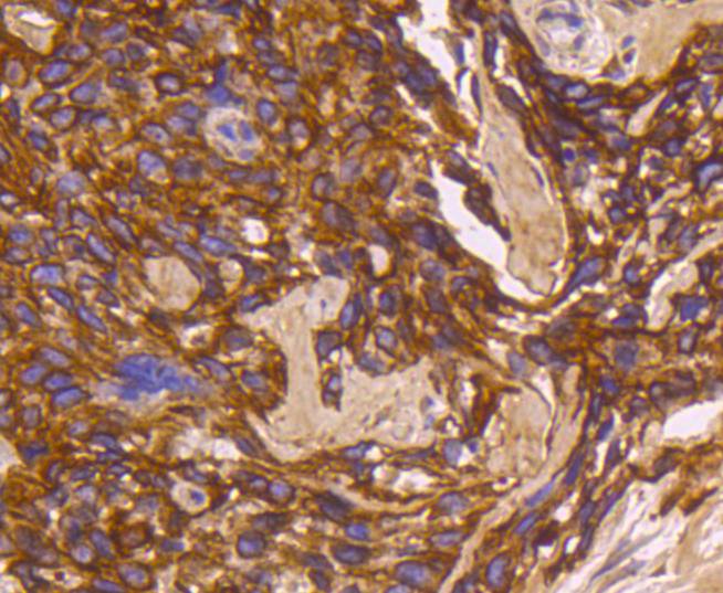
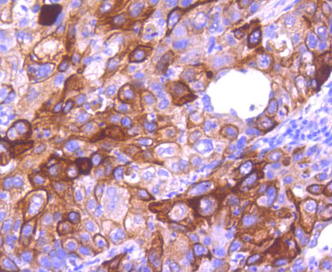
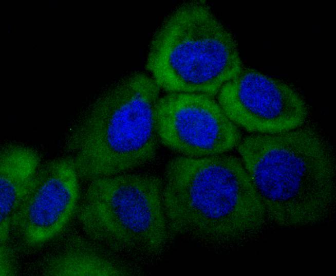
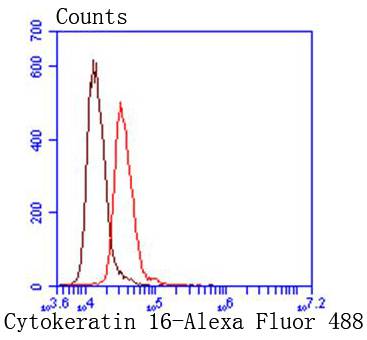
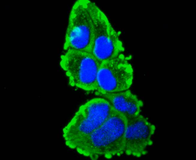
| 產(chǎn)品圖片 | Western blot analysis of Cytokeratin 16 on human skin tissue lysates. Proteins were transferred to a PVDF membrane and blocked with 5% BSA in PBS for 1 hour at room temperature. The primary antibody (bsm-52056R, 1/500) was used in 5% BSA at room temperature for 2 hours. Goat Anti-Rabbit IgG - HRP Secondary Antibody (HA1001) at 1:5,000 dilution was used for 1 hour at room temperature. Immunofluorescence analysis of paraffin-embedded human esophagus tissue labeling Cytokeratin 16 (bsm-52056R) and Vimentin (EM0401).The section was pre-treated using heat mediated antigen retrieval with Tris-EDTA buffer (pH 9.0) for 20 minutes. The tissues were blocked in 10% negative goat serum for 1 hour at room temperature, washed with PBS. And then probed with the primary antibodies Cytokeratin 16 (bsm-52056R, red) at 1/200 dilution and Vimentin (EM0401, green) at 1/400 dilution at +4℃ overnight, washed with PBS. Immunofluorescence analysis of paraffin-embedded human breast tissue labeling Cytokeratin 16 with Rabbit anti-Cytokeratin 16 antibody (bsm-52056R) at 1/200 dilution.The section was pre-treated using heat mediated antigen retrieval with Tris-EDTA buffer (pH 9.0) for 20 minutes. The tissues were blocked in 10% negative goat serum for 1 hour at room temperature, washed with PBS, and then probed with the primary antibody (bsm-52056R, red) at 1/200 dilution overnight at 4 ℃, washed with PBS. Immunohistochemical analysis of paraffin-embedded human breast carcinoma tissue using anti-Cytokeratin 16 antibody. The section was pre-treated using heat mediated antigen retrieval with Tris-EDTA buffer (pH 8.0-8.4) for 20 minutes.The tissues were blocked in 5% BSA for 30 minutes at room temperature, washed with ddH2O and PBS, and then probed with the primary antibody (bsm-52056R, 1/50) for 30 minutes at room temperature. The detection was performed using an HRP conjugated compact polymer system. DAB was used as the chromogen. Tissues were counterstained with hematoxylin and mounted with DPX. Immunohistochemical analysis of paraffin-embedded human lung carcinoma tissue using anti-Cytokeratin 16 antibody. The section was pre-treated using heat mediated antigen retrieval with Tris-EDTA buffer (pH 8.0-8.4) for 20 minutes.The tissues were blocked in 5% BSA for 30 minutes at room temperature, washed with ddH2O and PBS, and then probed with the primary antibody (bsm-52056R, 1/50) for 30 minutes at room temperature. The detection was performed using an HRP conjugated compact polymer system. DAB was used as the chromogen. Tissues were counterstained with hematoxylin and mounted with DPX. ICC staining of Cytokeratin 16 in A431 cells (green). Formalin fixed cells were permeabilized with 0.1% Triton X-100 in TBS for 10 minutes at room temperature and blocked with 1% Blocker BSA for 15 minutes at room temperature. Cells were probed with the primary antibody (bsm-52056R, 1/50) for 1 hour at room temperature, washed with PBS. Alexa Fluor?488 Goat anti-Rabbit IgG was used as the secondary antibody at 1/1,000 dilution. The nuclear counter stain is DAPI (blue). ICC staining of Cytokeratin 16 in HepG2 cells (green). Formalin fixed cells were permeabilized with 0.1% Triton X-100 in TBS for 10 minutes at room temperature and blocked with 1% Blocker BSA for 15 minutes at room temperature. Cells were probed with the primary antibody (bsm-52056R, 1/50) for 1 hour at room temperature, washed with PBS. Alexa Fluor?488 Goat anti-Rabbit IgG was used as the secondary antibody at 1/1,000 dilution. The nuclear counter stain is DAPI (blue). ICC staining of Cytokeratin 16 in Hela cells (green). Formalin fixed cells were permeabilized with 0.1% Triton X-100 in TBS for 10 minutes at room temperature and blocked with 1% Blocker BSA for 15 minutes at room temperature. Cells were probed with the primary antibody (bsm-52056R, 1/50) for 1 hour at room temperature, washed with PBS. Alexa Fluor?488 Goat anti-Rabbit IgG was used as the secondary antibody at 1/1,000 dilution. The nuclear counter stain is DAPI (blue). |
我要詢價(jià)
*聯(lián)系方式:
(可以是QQ、MSN、電子郵箱、電話等,您的聯(lián)系方式不會被公開)
*內(nèi)容:


